
A diagnosis of arthritis doesn't mean you have to stop doing the things you love. In fact, one of the most effective ways to manage arthritis is to keep moving.
While many people worry that exercise will make their joints worse, the opposite is often true. The right type of exercise can reduce pain, improve mobility, build strength, and help you stay independent for longer.
Working with an Accredited Exercise Physiologist (AEP) ensures your exercise programme is tailored to your individual needs, goals, and symptoms, giving you the confidence to move safely and effectively.
What Is Arthritis?
Arthritis is a broad term that describes conditions affecting the joints. The two most common types are:
Osteoarthritis (OA): The most common form of arthritis, caused by changes in joint cartilage and surrounding tissues over time. It most commonly affects the knees, hips, hands, and spine.
Rheumatoid Arthritis (RA): An autoimmune condition where the immune system attacks the joints, leading to pain, inflammation, and stiffness.
Although these conditions have different causes, both can lead to pain, reduced mobility, and difficulty performing everyday activities.
Common Symptoms of Arthritis
Arthritis can affect everyone differently, but common symptoms include:
Joint pain
Morning stiffness
Swelling around the joints
Reduced flexibility
Muscle weakness
Difficulty walking, climbing stairs, or getting up from a chair
These symptoms can gradually reduce confidence in movement, causing many people to become less active. Unfortunately, less movement often leads to weaker muscles, stiffer joints, and worsening symptoms.
Why Exercise Is So Important
Exercise is now recognised as one of the most effective non-surgical treatments for arthritis.
Regular physical activity can help:
Reduce joint pain
Improve mobility and flexibility
Increase muscle strength to better support the joints
Improve balance and stability
Reduce fatigue
Support weight management
Improve mood and overall wellbeing
Help maintain independence
The key is finding the right exercises and progressing them appropriately—not avoiding movement altogether.
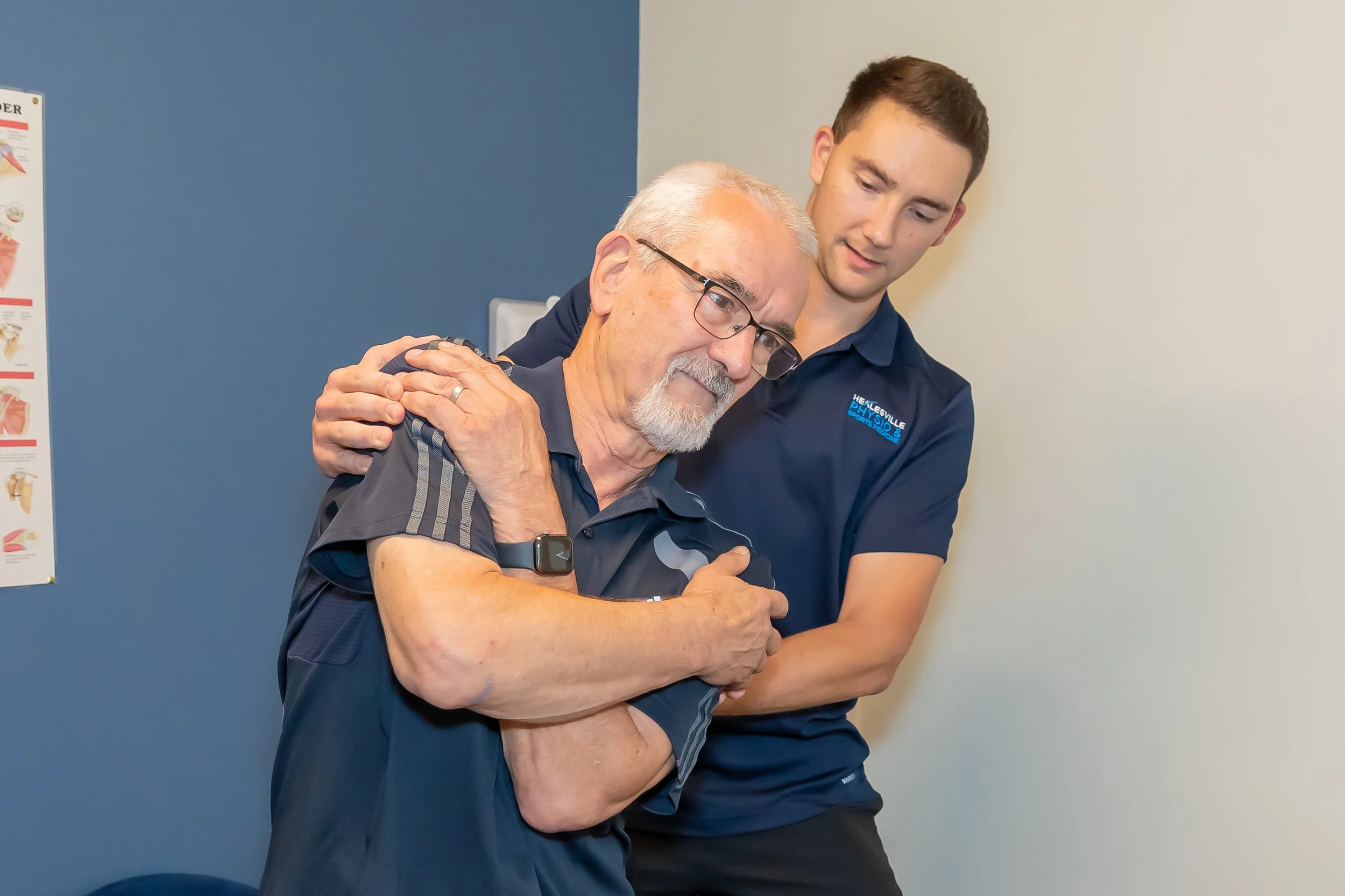
How an Exercise Physiologist Can Help
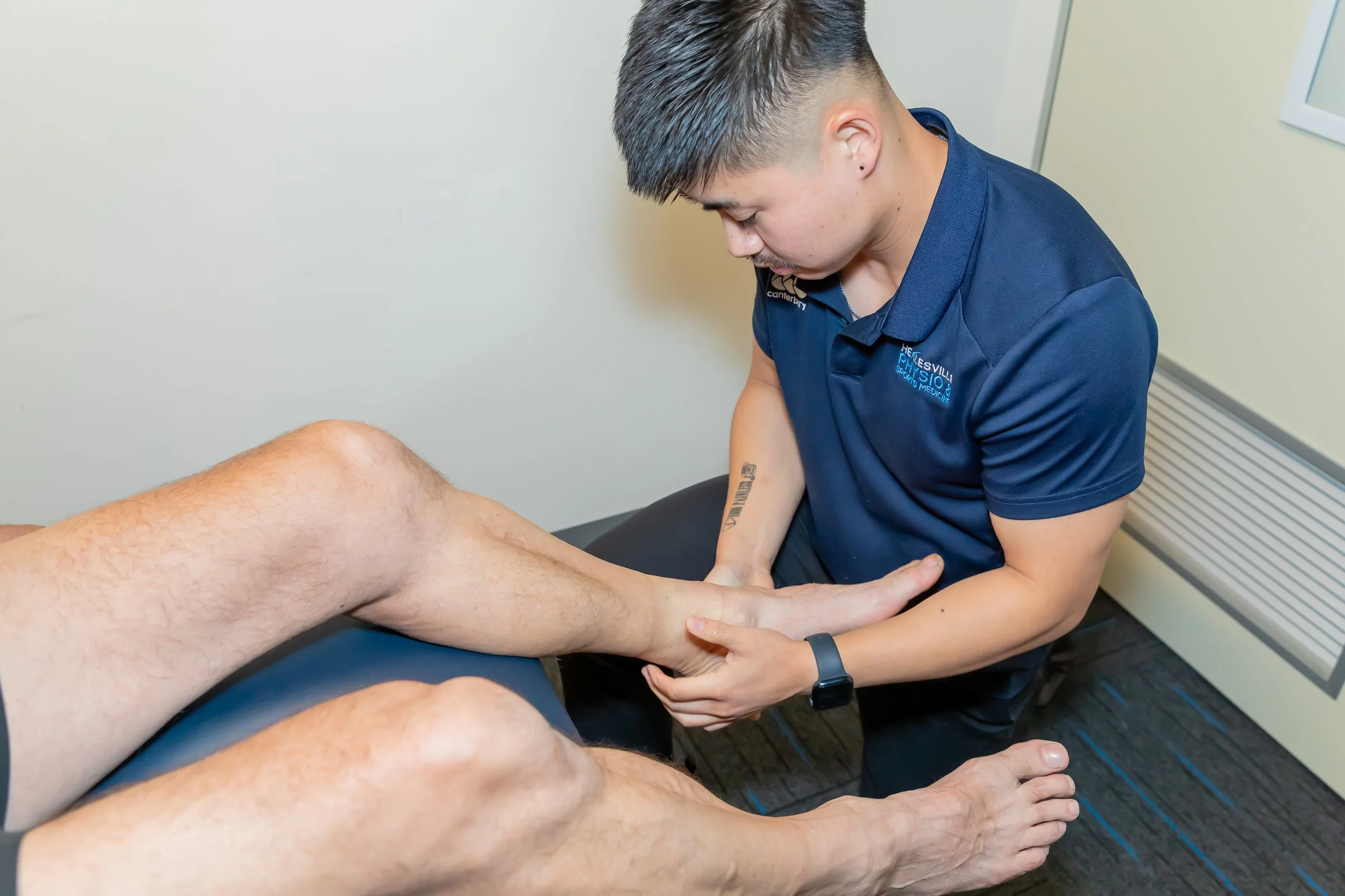
An Accredited Exercise Physiologist specialises in using exercise to manage chronic health conditions, including arthritis.
After assessing your mobility, strength, balance, pain levels, and goals, they'll develop an individualised exercise programme that is safe, achievable, and designed specifically for you.
Your programme may include:
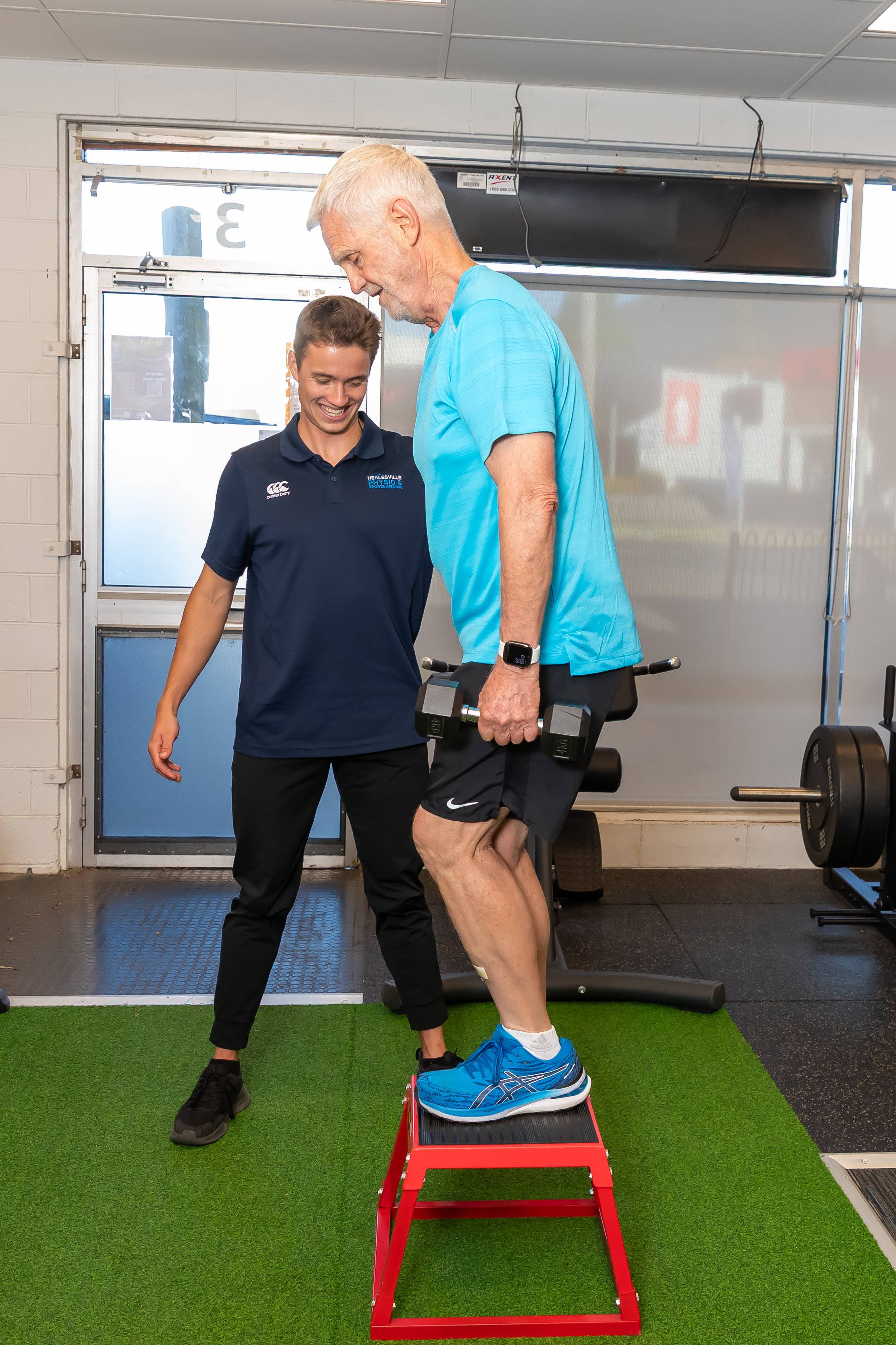
Strength Training
Stronger muscles provide better support for painful joints, helping to reduce discomfort during everyday activities.
Mobility and Flexibility Exercises
Gentle movement can improve joint range of motion and reduce stiffness, making everyday tasks easier.
Balance Training
Improving balance can reduce the risk of falls and increase confidence when moving around.
Aerobic Exercise
Low-impact activities such as walking, cycling, or swimming can improve cardiovascular health, reduce fatigue, and help manage body weight.
Education and Self-Management
An exercise physiologist can help you understand how arthritis affects your body, how to exercise safely during flare-ups, and how to build long-term habits that support joint health.
Introducing Our Osteoarthritis Exercise Classes
If you're living with osteoarthritis, exercising in a supportive and supervised environment can make all the difference.
At our clinic, we offer Osteoarthritis Exercise Classes led by an Accredited Exercise Physiologist. These small-group classes are specifically designed for people with osteoarthritis and focus on improving strength, mobility, balance, and confidence.
Each class includes exercises that can be modified to suit your individual ability, allowing you to work at your own pace while receiving professional guidance throughout the session.
Our classes provide more than just exercise—they're an opportunity to build confidence, stay motivated, and connect with others who are working towards similar goals.
Whether you're newly diagnosed or have been managing arthritis for years, these classes can help you stay active and improve your quality of life.
Everyday Tips for Managing Arthritis
Alongside regular exercise, these simple strategies can help manage symptoms:
Stay as active as possible.
Aim for regular movement throughout the day rather than long periods of sitting.
Choose low-impact activities that you enjoy.
Build strength gradually.
Maintain a healthy body weight to reduce stress on your joints.
Warm up before exercise.
Listen to your body and adjust your activity during flare-ups, but avoid complete rest where possible.
Consistency is far more important than intensity.
When Should You See an Exercise Physiologist?
You may benefit from seeing an Accredited Exercise Physiologist if you:
Have ongoing joint pain or stiffness
Have been diagnosed with osteoarthritis or rheumatoid arthritis
Want to exercise safely but aren't sure where to start
Have stopped being active because of pain
Want to improve your strength, mobility, and confidence
The earlier you begin an appropriate exercise programme, the better your chances of maintaining function and staying active.
Take the First Step Towards Better Joint Health
Living with arthritis doesn't mean accepting pain or giving up the activities you enjoy. With the right guidance, exercise can be one of the most powerful tools for managing symptoms and improving your quality of life.
Whether you're looking for a personalised exercise programme or would like to join one of our Osteoarthritis Exercise Classes, our Accredited Exercise Physiologist is here to help.
Get in touch with our team today to learn more about how exercise can help you move more comfortably, stay stronger, and continue doing the things that matter most.